The Gold Paper Award
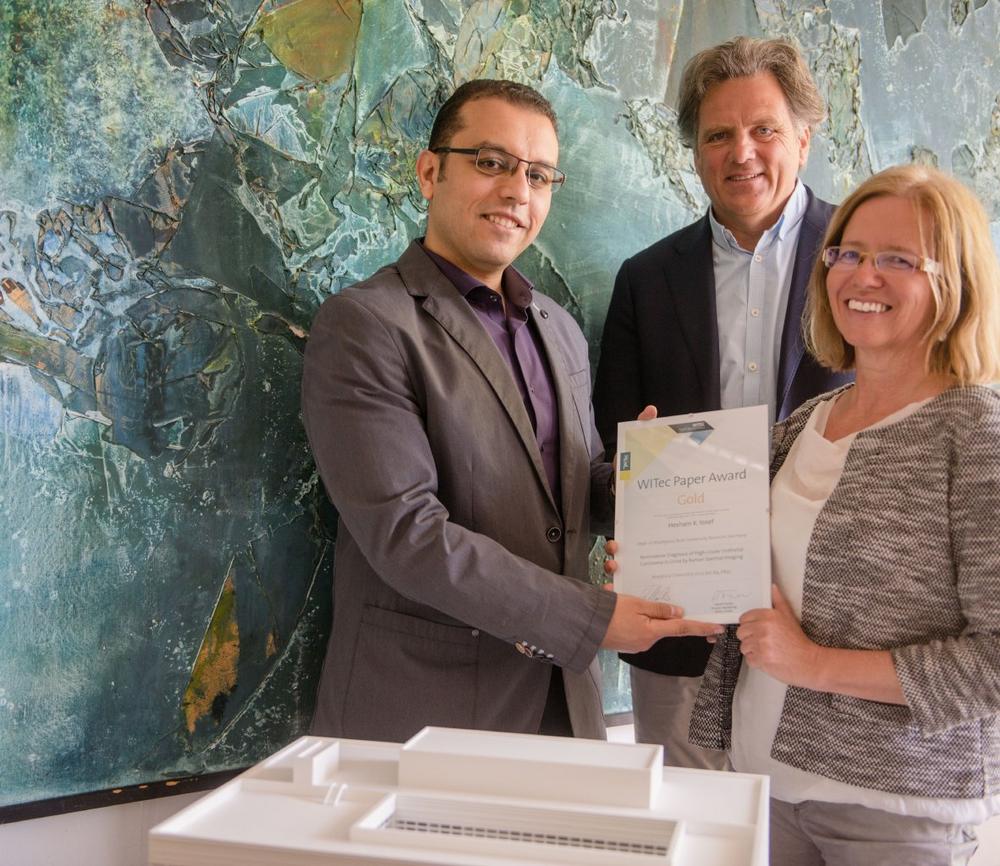
The Gold Paper Award goes to Hesham K. Yosef. With his colleagues from the Department of Biophysics, headed by Klaus Gerwert, and coworkers from other institutes at Ruhr-University Bochum (Germany) he developed a new, non-invasive approach based on Raman imaging for the detection of urothelial carcinoma cells in urine. Urothelial carcinomas constitute the majority of bladder tumors. Noninvasive, painless methods are urgently wanted for the diagnosis of this cancer and the monitoring of the treatment outcome. The scientists first identified differences in the Raman spectra of healthy and high-grade cancerous cells in urine. The most prominent differences in Raman bands indicated a decrease in the level of glycogen and an increase in the levels of fatty acids in the tumor cells, illustrating distinct metabolic changes. The scientists then developed and trained a classifier that could differentiate healthy from cancerous urothelial cells by their Raman spectra. A Raman band at 482 relative wavenumbers, indicating a massive loss of glycogen in tumor cells, emerged as a highly reliable marker. A first validation test on the urine of ten diagnosed and ten healthy patients was performed and the classifier diagnosed all samples automatically with 100% accuracy. First author of the publication, Hesham K. Yosef, says that he is convinced that, “The results will pave the way for spectral, noninvasive cytology of urine using Raman microspectroscopy.”
The Silver Paper Award
Mimicking biological structures with interesting functional features is a thriving field of research. Marvin Gernhardt, Holger Schmalz, Seema Agarwal and coworkers at the Chair of Macromolecular Chemistry II, University of Bayreuth, and the Bavarian Polymer Institute (Germany) were inspired by biological fibers and developed new fibers with distinct structures. They produced the fibers by side-by-side electrospinning of a thermo-responsive polymer and methacrylate-based copolymer. By changing the spinning solvents they fabricated side-by-side and coaxial bead-on-string fibers and analyzed these in detail with correlative Raman imaging, atomic force microscopy and scanning electron microscopy. The scientists concluded: “The work opens the way for the construction of heterostructured fiber morphologies based on different polymer combinations, offering high potential for applications as actuators, smart textiles, water management and catalysis.”
The Bronze Paper Award
Guanglin Yu receives the Bronze Paper Award. He and his colleagues from the Departments of Mechanical Engineering and Biomedical Engineering at the University of Minnesota in Minneapolis (USA) analyzed, when and how ice crystals develop in cells during freezing. Cryopreservation is a common method used to store cells, however not all cells survive the procedure. It has been hypothesized that a high cooling rate leads to the death of cells because of intercellular ice formation. How that happens is still a matter of debate. The researchers used confocal Raman imaging to monitor the freezing response of lymphoblasts under various conditions during the cryopreservation process. Ice crystals, cell mass and protective substances could be easily imaged and identified by their Raman spectra. As a parameter of cell death, they used a Raman signal indicating the release of cytochrome c from mitochondria. The imaging study showed that only the formation of large intracellular ice crystals (> 2 μm) is deadly for the cells and that a mixture of glycerol and trehalose prevented cell death most efficiently at a cooling rate of 10°C per minute. “Raman spectromicroscopy provides a powerful tool for observing IIF [intracellular ice formation] and understanding its role in cell death during freezing, and enables the development, to our knowledge, of new and improved cell preservation protocols,” concluded the authors in their paper.
2018 WITec Paper Award Winners
GOLD: Hesham K. Yosef, Sascha D. Krauß, Tatjana Lechtonen, Hendrik Jütte, Andrea Tannapfel, Heiko U. Käfferlein Thomas Brüning, Florian Roghmann, Joachim Noldus, Axel Mosig, Samir F. El-Mashtoly and Klaus Gerwert: Noninvasive diagnosis of high-grade urothelial carcinoma in urine by Raman spectral imaging. Analytical Chemistry 89, 6893 (2017), DOI 10.1021/acs.analchem.7b01403.
SILVER: Marvin Gernhardt, Ling Peng, Matthias Burgard, Shaohua Jiang, Beate Förster, Holger Schmalz and Seema Agarwal: Tailoring the morphology of responsive bioinspired bicomponent fibers. Macromolecular Materials and Engineering 303, 1700248 (2017), DOI. 10.1002/mame.201700248.
BRONZE: Guanglin Yu, Yan Rou Yap, Kathryn Pollock and Allison Hubel: Characterization intracellular ice formation of lymphoblasts using low-temperature Raman spectroscopy. Biophysical Journal 112, 2653 (2017), DOI 10.1016/j.bpj.2017.05.009.
The WITec Paper Award is an annual contest sponsored by WITec and awarded to author(s) to recognize his or her outstanding peer-reviewed publication featuring results acquired with a WITec instrument. Scientists from all fields of application in both academia and industry are invited to submit their publications to papers@witec.de. The deadline for submissions for the next Paper Award is January 31st, 2019.
WITec is a leading German manufacturer of confocal and scanning-probe microscopes for state-of-the-art Raman, Atomic Force (AFM), Scanning Near-Field Optical Microscopy (SNOM) and developed the integrated Raman Imaging and Scanning Electron (RISE) microscopy. WITec’s headquarters is located in Ulm, Germany, where all WITec products are developed and produced. Branch offices in USA, Japan, Singapore, and Spain ensure a worldwide sales and support network. From the company’s founding in 1997, WITec has been distinguished by its innovative product portfolio and a microscope design that enables combinations of the various imaging techniques within one system. An exemplar of the company’s breakthrough development is the world’s first integrated Raman-AFM microscope. To this day, WITec’s confocal microscopes are unrivaled in sensitivity, resolution and imaging capabilities. Significant innovation awards such as the Achema Innovation Award 2015 for the fully automated apyron microscope and a Prism Award 2015 for RISE microscopy document WITec’s enduring success and innovative strength.
WITec GmbH
Lise-Meitner-Str. 6
89081 Ulm
Telefon: +49 (731) 14070-0
Telefax: +49 (731) 14070-200
https://Raman.oxinst.com
Technisches Marketing
E-Mail: karin.hollricher@witec.de
![]()